Rapp. Comm. int. Mer Médit., 37,2004
213
DISTRIBUTION OF TRACE METALS IN DIFFERENT TISSUES OF BLUEFIN TUNA THUNNUS THYNNUS
Zorana Kljakovic-Gašpic*, Vjekoslav Ticina, Nikša Odžak, Tomislav Zvonaric, Ante Baric
Institute of Oceanography and Fisheries, 21 000 Split, Croatia - * kljakovic@izor.hr
Abstract
Concentrations of Cd, Cr, Cu and Zn were analyzed in muscle tissue, gill and liver samples of the bluefin tuna Thunnus thynnuscaught in
the Middle Adriatic. Analytical results revealed variable distribution of metals in the examined tissues. Maximum levels of Cd, Zn and Cu
were determined in the liver, while Cr concentrations were similar in all analyzed tissues.
Keywords : trace metals, distribution, bluefin tuna
Introduction
Data on trace metal content in different fish tissues are often used
to study the physiological behavior of metals in the fish (1). The aim
of this study was to determine the concentrations of some potentially
toxic trace metals (Cd, Cr, Cu and Zn) in different tissues of the
bluefin tuna Thunnus thynnus, in order to determine distribution
patterns of metals in the organism.
Methods
Eighteen specimens of bluefin tuna were caught by purse-seine in
the open waters of the Middle Adriatic, during August 1996. Collected
fish were immediately frozen (-20şC) and transported to the
laboratory for analysis. In the laboratory, the fork length (range: 123-
240 cm; mean=157±25 cm) and weight (range: 35-165 kg;
mean=69±27 kg) of each specimen were measured. Liver, gills, parts
of light muscles near the head, from the middle part and the tail of the
fish, as well as the red muscle from the middle part were cut out and
frozen prior to analysis. Preparation of tissues for trace metal analysis
included freeze drying, sample homogenization and wet digestion (2).
Trace metal analyses were performed using graphite furnace atomic
absorption spectroscopy. All results are reported in mg kg
-1
dry
weight. The accuracy of the analytical procedure was tested using
certified reference material IAEA-350 (Tuna fish). Statistical
differences between mean metal concentrations in different tissues
were evaluated using non-parametric Sign Test.
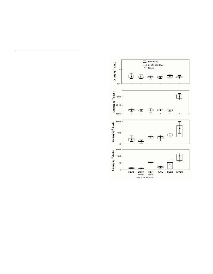
Results and discussion
Heavy metal concentrations in the different fish tissues are shown
in Fig. 1. Chromium concentrations were similar in all tissues
(P>0.05). In contrast, the highest levels of Cd, Zn and Cu were
determined in liver samples. Cadmium concentrations in liver were
45-55 times higher in comparison to all other tissues, among which
there were no significant differences (P>0.05). Concentrations of Zn
and Cu in liver were also 4-13 times and 9-66 times higher
respectively in comparison to other tissues. Observed patterns of trace
metal distribution between tissues match well with the results of other
field and laboratory studies (1, 3-5). Differences between trace metal
concentrations in analyzed tissues probably originate from differences
in physiological functions of muscles, gills and liver (1, 6). However,
distribution of Cu and Zn among the different muscle tissues also
differed. Concentrations of Zn in red muscle samples and in muscles
from near the head and the tail were higher than in the light muscle
samples from the middle part of the fish. Unlike Zn, Cu
concentrations were similar in the light muscle samples from three
different parts of the fish, and 5-8 times lower than in the red muscle.
This is probably due to differences in distribution and detoxification
strategies of Cu and Zn in fish (1). Higher Cu concentrations in red
muscle could also be related to the important metabolic role of Cu in
respiratory pigments (1), which are present at high concentrations in
the blood and the red muscle itself (6).
References
1-Phillips D.J.H., and Rainbow P.S., 1993. Biomonitoring of Trace
Aquatic Contaminants, Pp. 371. Elsevier Science Publ. LTD, Barking,
Essex.
2-Kljakovic-Gašpic Z., Zvonaric T., Vrgoc N., Odžak N., and Baric A.,
2002. Cadmium and lead in selected tissues of two commercially
important fish species from the Adriatic Sea. Water Res.,36: 5023-5028.
3-Odžak N., and Zvonaric T., 1995. Cadmium and lead uptake from food
by the fish Dicentrarchus labrax. Water Sci. Technol.,32: 49-55.
4-Širovic A., 1995. Correlation between weight of juvenile bluefin tuna
from the Adriatic and accumulation of lead and cadmium to its tissues. Pp.
32. International baccalaureate, XV Gymnasium, Zagreb.
5-Storelli M.M., Ceci E., and Marcotrigiano G.O., 1998. Comparative
study of heavy metal residues in some tissues of the fish Galeus
melastomus caught along the Italian and Albanian coasts. Rapp. Comm.
int. Mer Médit., 35(2): 288-289.
6-Bond C.E., 1996. Biology of fishes, 2nd ed., 750 p. Saunders College
Publ., Florida.
Fig. 1. Trace metal concentrations (in mg kg
-1
dry weight) in liver, gills
and muscle tissues of bluefin tuna (N=18; length: 123-240 cm; weight:
35-165 kg). Dots represent average values; lower and upper box edges
represent average ±1 SD; outlying bars are minimum and maximum
values.