Rapp. Comm. int. Mer Médit., 37,2004
251
METAL CYCLING THROUGH PLANKTON COMMUNITIES: A SINGLE-CELL APPROACH
USING SYNCHROTRON-BASED X-RAY FLUORESCENCE
Benjamin S. Twining†, Stephen B. Baines, and Nicholas S. Fisher*
Marine Sciences Research Center, State University of New York, Stony Brook, NY 11794-5000 USA - * nfisher@notes.cc.sunysb.edu
†Present address: Yale School of Forestry and Environmental Studies,
Environmental Science Center, PO Box 208105, New Haven, CT 06520
Abstract
We have applied a synchrotron-based x-ray ?uorescence microprobe to analyze the concentrations and cytological distributions of trace
elements in autotrophic and heterotrophic protists from remote waters. Using this approach it is now possible to discern different elemental
stoichiometries exhibited by different types of co-occurring protists. This technique, combined with other geochemical measurements,
should enable important new advances in our understanding of the marine biogeochemistry of trace elements.
Keywords: x-ray ?uorescence, microprobe, microbial loop, trace metals
Introduction
The elemental composition of marine protists is of great interest to
oceanographers. The elemental stoichiometries of plankton
simultaneously re?ect the nutrient ratios of the aquatic environment
and control the input of recycled elements through remineralization of
plankton (1). While most attention has been focused on the C, N, and
P content of plankton, several early studies determined the trace metal
composition of plankton as well (2, 3). More recent evidence that
trace metal nutrients such as iron can limit primary production in both
open-ocean and coastal environments (4, 5) has spurred researchers to
further study the trace metal contents of plankton.
Bioactive trace metals such as Mn, Fe, Co, Ni, Cu, and Zn are
typically measured in plankton by first concentrating cells onto filter
membranes and then digesting the filters and analyzing the resulting
solution with atomic absorption spectrometry or inductively-coupled
plasma mass spectrometry. This approach requires concentrating large
amounts of similarly sized cellular and abiotic material on membranes
of various pore-size. Thus co-occurring plankton with overlapping
size ranges cannot be separated, and the potentially contaminating
in?uence of suspended abiotic particles cannot be eliminated.
We have developed a new approach to the analysis of trace
elements in marine protists that utilizes the unique sensitivity of
synchrotron x-ray radiation to measure trace metals in individual
nanoplankton cells. Further, the synchrotron x-ray ?uorescence
(SXRF) technique produces a two-dimensional map of the metals in
each cell, providing additional information on the co-localization of
elements. Here we present an example of the information that can be
collected using this technique for cells collected from the Southern
Ocean during a recent mesoscale iron enrichment experiment
(SOFeX).
Materials and methods
Complete descriptions of the sample preparation and analysis
protocols are presented elsewhere (6). Brie?y, cells were collected
with trace-metal ‘clean’techniques from the Southern Ocean, before
and after Fe fertilization, and immediately centrifuged onto gold
electron microscopy grids following fixation with Chelexed
glutaraldehyde. The mounted cells were brie?y rinsed with Milli-Q
deionized water and then dried in a Class 100 laminar ?ow hood.
Light and epi?uorescence micrographs of the dried cells were taken
on-board the ship, and the cells stored in a plastic dessicator until
analysis. SXRF analyses were performed at the 2-ID-E beamline of
the Advanced Photon Source, Argonne National Laboratory, Argonne,
IL. Each cell was raster scanned across a highly focused x-ray spot,
and the excited x-ray ?uorescence spectra were recorded at each pixel
with a 3-element germanium detector. Spectra were averaged over the
cell, corrected for ?uorescence from nearby background regions, and
the peak areas modeled. Peak areas were converted to element
concentration with NIST thin-film standards. Trace element contents
were normalized to cellular P, which was measured directly with
SXRF.
Results and discussion
SXRF enables the identification of different trace metal
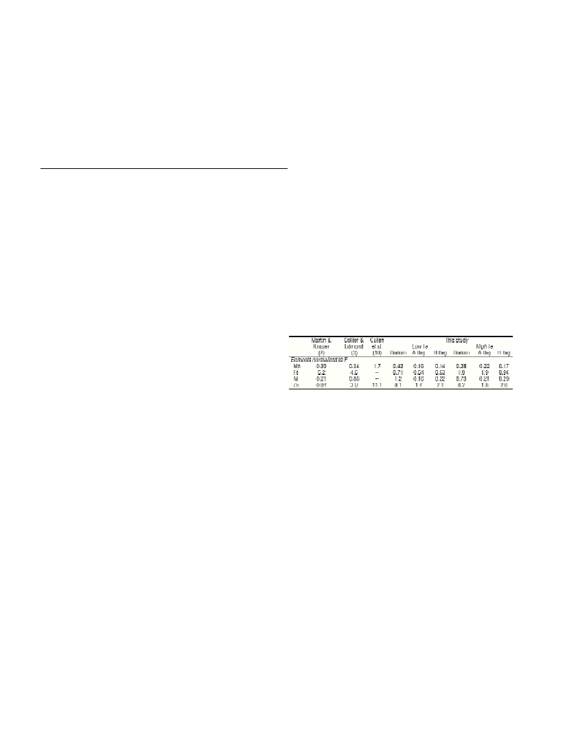
compositions of co-occurring protists cells. Table 1 presents an
example of SXRF data collected from the Southern Ocean, which are
compared to data from bulk analyses of collected plankton from other
waters. There are notable differences in the metal contents of diatoms
and ?agellated cells that cannot be detected with bulk analyses.
Flagellated cells were significantly more enriched in P and diatoms
more enriched in Mn, Ni, and Zn. Iron fertilization resulted in sharp
increases in cellular concentrations of Mn, Ni, and Zn and smaller
increases in P. Generally, P, Fe, and Zn were found distributed within
cells and Si in the frustules of diatoms. Adsorbed Fe, localized in high
concentrations attached to some cells in a way that doesn’t correspond
to any cellular feature, can be identified and removed from the
elemental analysis with SXRF. Explanations for varying elemental
stoichiometries in different co-occurring taxa remain to be discovered.
Stoichiometric variations among different taxa suggest that the
concept of a constant Redfield-type elemental ratio may not extend to
trace metals. Application of SXRF analyses to protists in other waters,
such as P-limited waters of the eastern Mediterranean, may reveal
different stoichiometric relationships and may help explain the
distribution of plankton in those waters.
Table 1. Elemental composition (ratios normalized to cellular P, mmol
mol
-1
) of natural plankton assemblages as measured with bulk analysis
and SXRF.
Shown are geometric mean stoichiometries for three cell types (diatoms,
autotrophic ?agellated cells—A ?ag, heterotrophic ?agellated cells—H ?ag)
collected from either low Fe (unenriched) or high Fe (enriched) stations. The
Martin and Knauer (2) and Collier and Edmond (3) data are shown as selected
by Bruland et al.(7). The SXRF Fe data are from Twining et al.(8, 9).
References
1-Redfield A.C., Ketchum B.H. and Richards F.A., 1963. The in?uence
of organisms on the composition of seawater. Pp. 26-77. In:M. N. Hill
(ed.), Comparative and Descriptive Oceanography. Wiley, New York.
2-Martin J.H. and Knauer G.A., 1973. The elemental composition of
plankton. Geochim. Cosmochim. Acta, 37: 1639-1653.
3. Collier R. and Edmond J., 1984. The trace-element geochemistry of
marine biogenic particulate matter. Prog. Oceanogr., 13: 113-199.
4-Martin J.H., Gordon R.M. and Fitzwater S.E., 1991. The case for iron.
Limnol. Oceanogr., 36: 1793-1802.
5-Hutchins D.A. and Bruland K.W., 1998. Iron-limited diatom growth
and Si:N uptake ratios in a coastal upwelling regime. Nature, 393: 561-
564.
6-Twining B.S., Baines S.B., Fisher N.S., Maser J., Vogt S., Jacobsen C.,
Tovar-Sanchez A. and Sanudo-Wilhelmy S.A., 2003. Quantifying trace
elements in individual aquatic protist cells with a synchrotron x-ray
?uorescence microprobe. Anal. Chem., 75: 3806-3816.
7-Bruland K.W., Donat J.R. and Hutchins D.A., 1991. Interactive
in?uences of bioactive trace-metals on biological production in oceanic
waters. Limnol. Oceanogr., 36: 1555-1577.
8-Twining B.S., Baines S.B. and Fisher N.S., 2004. Trace element
composition of individual plankton cells collected during the Southern
Ocean Iron Experiment (SOFeX). Limnol. Oceanogr., Submitted.
9-Twining B.S., Baines S.B., Fisher N.S. and Landry M.R., 2004.
Cellular iron contents of plankton during the Southern Ocean Iron
Experiment (SOFeX). Deep-Sea Res. I, Submitted.
10-Cullen J.T., Chase Z., Coale K.H., Fitzwater S.E. and Sherrell R.M.,
2003. Effect of iron limitation on the cadmium to phosphorus ratio of
natural phytoplankton assemblages from the Southern Ocean. Limnol.
Oceanogr., 48: 1079-1087.