REPRODUCTIVE BIOLOGY OF CHLOROPHTHALMUS AGASSIZI
IN THE CENTRAL-WESTERN MEDITERRANEAN
Follesa M.C.*, Cabiddu S., Davini M.A., Porcu C., Cau A.
Department of Animal Biology and Ecology, University of Cagliari, Italy - * follesac@unica.it
Abstract
160 specimens of Chlorophthalmus agassiziwith total length between 6.6 and 18.8 cm were caught in the Sardinian Sea. Microscopic
analysis showed six stages of oocyte development. Spawning occurred in Summer.
Key-words: Chlorophthalmus agassizi, hermaphroditism, reproduction, Mediterranean Sea
Rapp. Comm. int. Mer Médit., 37,2004
356
Introduction
Chlorophthalmus agassiziBonaparte, 1840,shortnose greeneye, is
a bathydemersal monoecious species (1). Though abundant in
Mediterranean Sea where it has a rather wide geographic distribution
(2, 3), the current knowledge on its biology is limited, especially on
its reproductive cycle. This paper studied its reproductive period.
Materials and methods
Samples were caught with experimental trawl surveys (Autumn and
Summer) carried out in the Sardinia Sea as part of national and
international (2) projects in 2002, and from samplings in the months
not covered by the experimental surveys. For each month, the gonads
of several individuals were collected stratified based on size.
Stages of oocyte development were based on those proposed by
Forberg (4) and adapted to the studied species. Oocytes were
measured and counted by stage and the Nucleoplasmatic Ratio (NPR)
was calculated: NPR=Vn(Vc-Vn)
-1
, where Vn = nucleus volume and
Vc = cytoplasm volume.
Results and discussion
The gonads were made up of two distinct components: the ovarian
and the testicular components. The ovarian component presented a
double sacciform structure elongated in the abdominal cavity. The
oviduct is one and gathers the mature sexual products of both ovaries.
The testicular portion, included in the median zone of each ovary, was
made up of seminiferous tubules and ended in an ampoule.
Following histological analysis of the gonads of 160 specimens
caught from March to November with a TL from 6.6 to 18.8 cm, six
oocyte development stages were identified (Fig. 1):
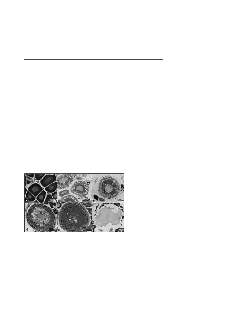
Fig. 1. Oocyte developmental stages: Basophils (a), Lipid vesicles (b),
Primary vitellogenesis (c), Secondary vitellogenesis (d), Tertiary vitello-
genesis (e), and Translucent (f).
B (Basophils): oocytes with a uniform cytoplasm, strongly
basophilic and homogeneous. NPR=0.211; diameter=20-70
µ
m (Fig.
1a).
LV (Lipidic Vesicles): oocytes with weakly basophilic cytoplasm,
which contained lipid vesicles. Follicular Cells (FC) and the “Zona
Radiata” (ZR) appeared. NPR=0.187; diameter=70-210
µ
m (Fig. 1b).
Y1 (Primaryvitellogenesis): the oocytes presented drops of protein
vitellin (Y=yolk) scattered in the cytoplasm and lipid vesicles
increased in number; the follicular cells started to thin out and the ZR
to thicken. NPR=0.056; diameter=210-270
µ
m (Fig. 1c).
Y2 (Secondaryvitellogenesis): the cytoplasm was acidophilous;
the drops of protein vitellin increased in number and size and started
to unite into progressively larger drops; the ZR appeared very thick
and the follicular cells continued to spread. NPR=0.021
diameter=270-500
µ
m (Fig. 1d).
Y3 (Tertiaryvitellogenesis): the drops of protein vitellin were
completely joined together as well as the lipid vesicles. The nucleus
migrated to the periphery of the oocyte until it disappeared
completely. Diameter >500
µ
m (Fig. 1e).
T (Translucent): the oocyte was large and characterised by
clarification of the protein vitellin. The oocyte was ready to be shed
(Fig. 1f).
Four stages of development have been assigned to the male portion
of the gonad: primary spermatocyte, secondary spermatocyte,
spermatid, and sperm.
Two periods of oocyte development were identified in a year: 1)
spent ovary, (an ovary with immature oocytes: B, LV); 2) ovary with
oocytes belonging to all the stages of maturation and sperms which
were also mature. The reproductive season started in May and ended
in September with a reproductive peak in July. In the mature ovaries
the contemporary presence of oocytes at progressive stages of
development was pointed out (asynchronous ovary), the eggs were
shed several times during a reproductive season.
It has been possible to hypothesise a simultaneous type of
hermaphroditism. During the reproductive season, in fact, the
presence of mature male and female elements was observed (TL>9
cm). The species seems to be sequentially protandric hermaphroditic:
very small individuals (TL 5-8 cm) presented immature ovaries during
all the months of the year, they always showed mature sperm.
Nevertheless the adopted reproductive strategy is still not clear.
Self-fertilisation could be possible for anatomical reasons (the male
and female genital tracts meet at one and the same opening),
moreover, the gonads contemporaneously present mature female and
male sexual elements. However, C. agassizilives in large shoals,
which suggests that meeting a sexual partner would not be difficult.
This hypothesis is further confirmed by the presence of a luminous
organ of symbiotic bacteria in the perianal area (5), which could be
used for sexual attraction, being an intraspecific system of
communication.
References
1-Merrett N.R., 1990. Chlorophthalmidae. Pp. 351-360. In: Quero J.C.,
Hureau C., Karrer A., Post and Saldanha L. (eds.), Check-list of the fishes
of the eastern tropical Atlantic (CLOFETA). JNICT, Lisbon; SEI, Paris;
and UNESCO, Paris.
2-Relini G., Bertrand J., and Zamponi A., (eds) 1998.Sintesi delle
conoscenze sulle risorse da pesca dei fondi del Mediterraneo centrale
(Italia e Corsica). Biol. Mar. Medit., 6 (suppl. 1): 165-168.
3-Sulak K.J., 1986. Chlorophthalmidae. Pp. 412-413. In:Whitehead P.J.,
Bauchot M.L., Hureau J.C., Nielsen J. and Tortonese E. (eds.), Check-list
of the fishes of the North-eastern Atlantic and the Mediterranean
(CLOFNAM). UNESCO, Paris; and ONU, Paris.
4-Forberg K.G., 1982. A histological study of development of oocytes in
capelin, Mallotus villosus villosus (Muller). J. Fish Biol., 20:143-154.
5-Somiya H., 1977. Bacterial bioluminescence in chlorophthalmid deep-
sea fish: a possible interrelationship between the light organ and the eyes.
Experentia, 33/7: 906-909.