PARASITIC DINOFLAGELLATES IN MEDITERRANEAN ZOOPLANKTON
Alf Skovgaard* and Enric Saiz
Institut de Cičncies del Mar (CMIMA - CSIC), Barcelona, Catalunya, Spain - * skovgaard@icm.csic.es, enric@icm.csic.es
Abstract
We investigated the occurrence of parasitic dino?agellates in zooplankton (copepods and appendicularians) off Barcelona, Spain. The most
common species were the intestinal parasites Blastodiniumspp. found in the copepod genera Oncaea, Corycaeus, Oithonaand
Paracalanus. However, also the coelomic parasite Syndinium was relatively common in Paracalanus. The ectoparasite Oodinium pouchetii
occasionally infested large proportions of appendicularians.
Keywords: Dino?agellate, Parasite, Copepod, Blastodinium, Syndinium
Rapp. Comm. int. Mer Médit., 37,2004
441
A number of dino?agellates are parasites of marine zooplankton
organisms (1) and, at times, these parasites may contribute
significantly to the mortality of copepod populations (2, 3). However,
the profusion of parasitism and its impact on zooplankton population
dynamics has received little attention and is not very well understood.
To study the prevalence and possible impact of dino?agellate
parasites, we collected zooplankton weekly or biweekly off Port
Olímpic, Barcelona, by taking vertical net tows with 53 and 100 µm
plankton nets. Live animal were observed qualitatively within 2-5
hours after sampling and, in addition, samples fixed in formaldehyde
were studied quantitatively for the prevalence of parasitized
zooplankton organisms.
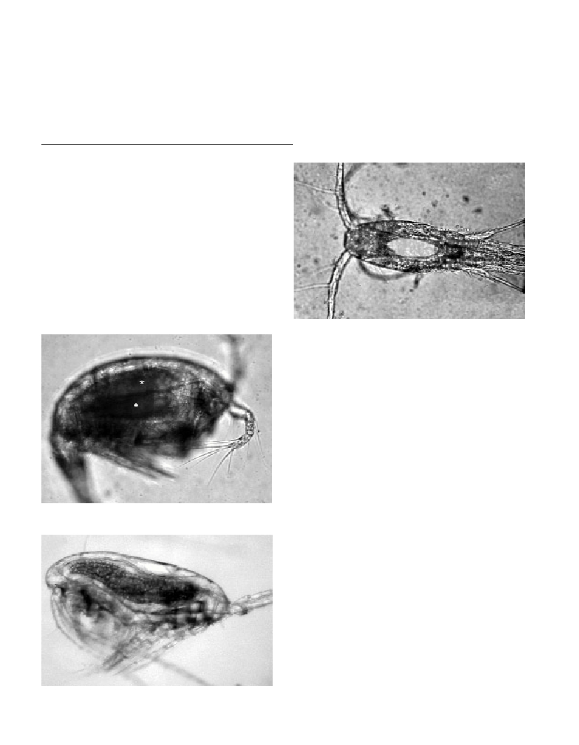
Blastodiniumspp. could be detected in live copepods as large
greenish or brownish bodies in the gut of infected animals (Figs 1-3).
In fixed animals, infection with Blastodinium was less evident due to
the lack of colour as a diagnostic tool. Based on the host species and
the size and shape of the parasites (1), we were able to identify at least
3 Blastodiniumspecies: B. manginiChatton (including the variety B.
manginivar. oncaeaChatton), B. contortumChatton and B. oviforme
Chatton. Syndinium turbo Chatton could be detected equally well in
live and fixed animals (P. parvus) since this parasite made its host
conspicuously dilated, dark and opaque (2, 3). The detection of
Oodiniumon appendicularians was straightforward, since these are
ectoparasites and form characteristic cysts attached the epithelium of
the host. When infected copepods were kept in seawater in the
laboratory, sporulation always occurred within 1-2 days and free-
swimming parasite zoospores could then be observed. This indicates
that only the final developmental stages of the parasite life cycles
within its copepod hosts were detected. New methods, therefore, need
to be applied in order to gain more knowledge on the total infection
frequencies and, thereby, the effect of dino?agellate parasites on their
copepod hosts.
References
1-Chatton, Č., 1920. Les Péridiniens parasites: morphologie, repro-
duction, éthologie. Arch. Zool. Exp. Gen., 59: 1-475.
2-Ianora, A., Scotto di Carlo, B., Mazzocchi, M. G., and Mascellaro, P.,
1990. Histomorhological changes in the reproductive condition of
parasitized marine planktonic copepods. J. Plank. Res., 12: 249-258.
3-Kimmerer, W. J., and McKinnon, A. D., 1990. High mortality in a
copepod population caused by a parasitic dino?agellate. Mar. Biol., 107:
449-452.
Fig. 1. Oncaeasp. female infected with two individuals of Blastodinium
mangini var. oncaea(asterisks). Live specimen.
Fig. 2. Paracalanus parvusfemale infected with Blastodinium contor-
tum.Live specimen.
Fig. 3. Oithona nanainfected with Blastodinium oviforme.Live specimen
viewed in eip?uorescent light. Fluorescence of Blastodiniumchlorophyll
is seen as bright ovoid body.