INITIAL PHASE OF THE INTRAOVARIAN SPERM STORAGE OF HELICOLENUS DACTYLOPTERUS
Sílvia Vila*, Maria Sŕbat, Marta Muńoz and Margarida Casadevall
Ŕrea de Zoologia, Departament de Cičncies Ambientals, Universitat de Girona, Campus Montilivi, Girona, Spain
* silvia.vila@udg.es, maria.sabat@udg.es, marta.munyoz@udg.es, margarida.casadevall@udg.es
Abstract
Sperm storage crypts located within the ovaries of Helicolenus dactylopterus were analysed during the first phase of this storage. Electron
and optical microscope observations showed some degree of organization of the stored spermatozoa. Besides, desmosomic unions among
cryptal cells denoted the isolation of spermatozoa inside the crypts and were probably related to the protection of these cells from the
immune system of the female.The retainment of an important portion of cytoplasm by the spermatozoa at the beginning of the storage
could be explained as an initial source of nutrients while there is not nutritional supply from the female.
Key words: Helicolenus dactylopterus, sperm storage, histology, ultrastructure
Rapp. Comm. int. Mer Médit., 37,2004
456
The bluemouth rockfish, Helicolenus dactylopterus, is a zygo-
parous oviparous species with internal fertilization. Females present
specialized structures known as crypts where the spermatozoa are
stored and mantained viable for periods up to 10 months, from April
and May onwards. When spawning starts, between January and
February, the spermatozoa stored in the crypts must be reactivated,
with subsequent fertilization of mature eggs (1).
We found that Helicolenusdacctylopterusshows the most complex
structures for sperm storage than those previously described in
viviparous species within the same family (2-3). Thus, in this species
spermatozoa remain grouped within differentiated structures instead
of ?oating freely like in Alcichthysalcicornis (3) or singly adhered to
the ovarian epithelium as found in viviparous species like Sebastes
taczanowskiithat retain spermatozoa adhered to the epithelium of the
ovigerous lamella or wrapped in its microvilli. The prolonged time
period during which spermatozoa must reside in the ovary may offer
an explanation about the existence of these specialised storage crypts.
The aim of this paper is to analyze the structure and ultrastructure
of these intraovarian crypts during the initial period of storage. From
June onwards there is a marked increment of testicular activity (4),
thus our work has been focused on two fresh samples caught in July
(Palamós, Costa Brava, northwest Mediterranean), when the crypts
are full of “new” spermatozoa.
In both cases several portions of the central ovarian area were
analysed. Small pieces of ovary were fixed in glutaraldehyde (2.5%)-
paraformaldehyde (2%) mixture in a 0.1 M cacodylate buffer.
Spermatozoa storage structures are located very near the muscular-
connective rachis of the gonad, at the base of the interlamellar gaps
(5). The cryptal epithelial cells present a nucleus with some strongly
condensed heterochromatin, a lot of free ribosomes, vesicles and
RER, and this is a clear evidence of important protein synthesis.
We also observed an enormous quantity of desmosomal unions
among the cryptal cells (Fig. 1), forming thus a continuous cellular
layer that surrounds the crypt cavity, probably related to the protection
of the spermatozoa from the immune system of the female which
stores them (3). Lining the epithelial cells there are some cells of
different morphology, with a nucleus slightly extended and much
more electrodense.
Our observations through the optical microscope suggested some
organization of the male gametes located in the crypts, a fact that later
was corroborated by the transmission electronic microscope. We can
clearly see that these organizations are not due to the existence of a
separate membrane like the ones which surround spermato-
zeugmata.(6), but the spermatozoa are arranged in some kind of bun-
dles seemingly independent one from one another.
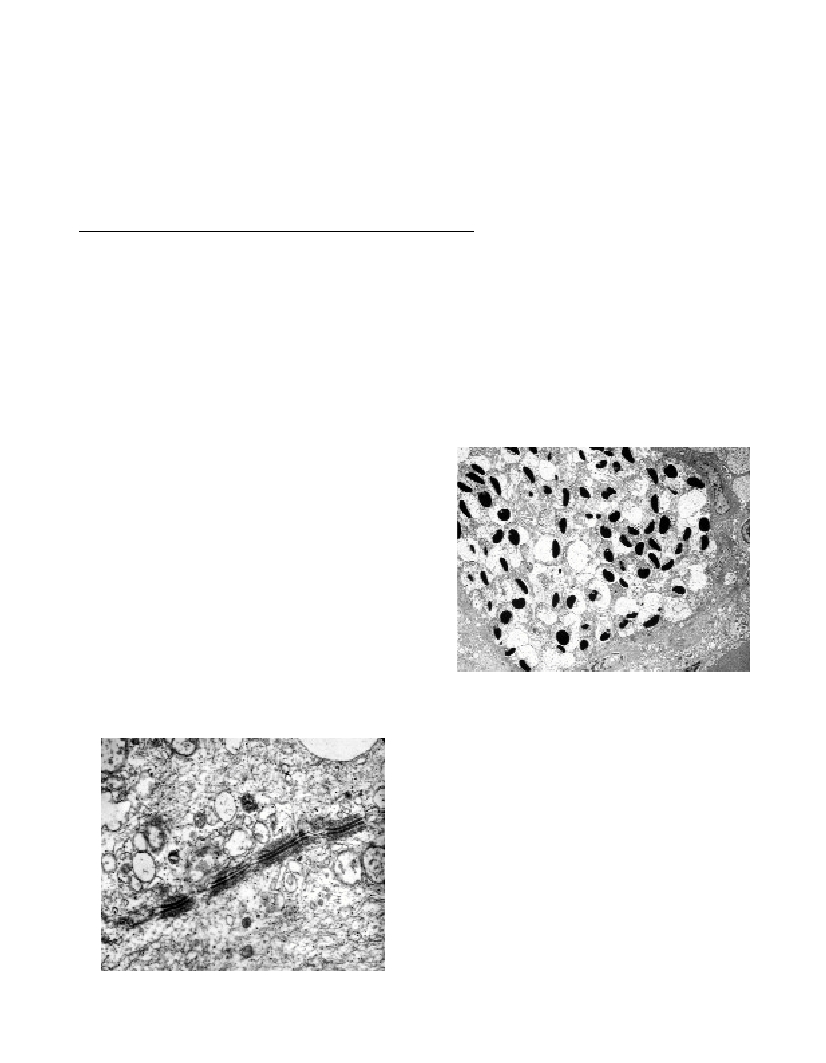
An outstanding fact is that the spermatozoa remaining inside the
crypts keep an important portion of cytoplasm (Fig. 2), in which we
can observe many mitochondria, vacuola and tiny granules. A possible
explanation could be that this cytoplasm represents an initial reserve
of nutrients for spermatozoa at the beginning of the storage period,
while later there would be evidence of a supply of nutritional
substances from the female towards the male sexual cells, a fact that
we aim to confirm in further studies.
References
1-Muńoz M., Casadevall M., Bonet S.,1999. Annual reproductive cycle
of Helicolenus dactylopterus dactylopterus (Teleostei: Scorpaeniformes)
with special reference to the ovaries sperm storage. J. Mar. Biol. Ass. UK.
79:521-529.
2-Moser, H.G. 1967. Seasonal histological changes in the gonads of
SebastodespaucispinisAyres, an ovoviviparous teleost (family
Scorpaenidae). J. Morphol. 123: 329-354.
3-Koya Y., Munehara H.,and Takano K., 1997. Sperm storage and
degradation in the ovary of a marine copulating sculpin, Alcichthys
alcicornis(Teleostei: Scorpaeniformes): role of intercellular junctions
between inner ovarian epithelial cells. JMorphol. 233: 153-163.
4-Muńoz, M. and M. Casadevall 2002. Reproductive indices and
fecundity of Helicolenus dactylopterus dactylopterus(Teleostei:
Scorpaenidae) in the Catalan Sea (western Mediterranean). J. Mar. Biol.
Ass. U.K. 82: 995-1000.
5-Muńoz M., Casadevall M., Bonet S.,and Quagio-Grassiotto I., 2000.
Sperm storage structures in the ovary of Helicolenus dactylopterus
dactylopterus(Teleostei: Scorpaeniformes): an ultrastructural study.
Environmental Biology of Fishes. Kluwer Academic Publ., Netherlands.
58: 53-59.
6-Downing, A. L. and J. R. Burns (1995). Testis morphology and
spermatozeugma formation in three genera of viviparous halfbeaks:
Nomorhamphus, Dermogenys, andHemirhamphodon (Teleostei:
Hemiramphidae). Journal of Morphology. 225: 329-343.
Fig. 1. Transmission electron micrograph showing desmosomal unions
among cryptal epithelial cells.
Fig. 2. Intraovarian sperm storage crypts. Electron microscopy view
showing a sperm bundle surrounded by cryptal epithelial cells.