Introduction
Based upon acute toxicity tests, mercury is important in bioaccu-
mulation and sublethal effects in marine organisms. In its ecological
cycle Hg is found in a part of the Earth’s crust, its water and atmos-
phere layer, but when incorporated in living organisms mercury may
perturb vital bio chemical processes. The fish that live in marine
waters could obtain their mercury load directly from the seawater and
through the food web.This accumulated mercury may increase with
fish age, and one such example is the tuna, Thunnus thynnusL. Since
the Mediterranean is considered to be an area of enhanced natural mer-
cury input (the biggest world mercury mines are situated on the edges
of this basin) (1), it is possible that tuna in the Adriatic contain signif-
icant mercury levels.Toxic effects of mercury are expressed in differ-
ent ways according to the chemical form of Hg, the dose, and the route
of exposure in various species of animals.
Selenium as a rare metalloid is both essential and toxic within a rel-
atively narrow concentration range. A biological interest in selenium
has significantly increased during the last four decades when its use-
ful in?uence in living organisms was established (2,3). The fact that
selenium is essential in the metabolic defence of an organism against
oxide stress (4) was an important discovery.
Materials and methods
The tuna were obtained by professional fishermen in middle
Adriatic area (around the Islands of Vis and Jabuka) in the summer of
1996. After biometric measurements (weight, length and age defining
of fish) were made, two organs (liver and gills) and two comestible tis-
sues (light and dark muscle) were taken for analysis. The samples
were homogenized, placed in polyethylene pots and stored in refriger-
ator at –20°C.
The quantitative analysis was carried out by A.A.S. (Perkin Elmer
1100B) with hydride generation after organic matrix digestion with
HNO
3
/H
2
O
2
for Hg and HNO
3
/H
2
SO
4
/HClO
4
/HCl for Se. The accu-
racy of the analytical procedures was tested and controlled using
NRCCTORT-1 (lobster hepatopancreas) and DORM-2 (dogfish mus-
cle) certified reference material. A comparison of the obtained results
with the reference values is shown in Table 1.
Table 1. Results of standard reference-material analysis.
The obtained results showed very good compliance with the refer-
ence values.
Results
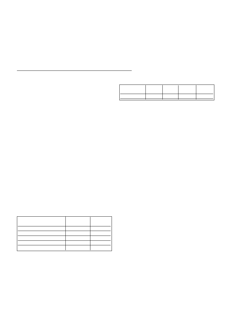
Measured average values of mercury and selenium concentrations
in the selected organs and tissues are shown in Table 2.
Table 2. Average values (±SD) of Hg and Se concentrations
(mg/kg wet weight) in the selected organs and tissues
Conclusions
The results obtained lead to the following conclusions:
- It is determined that the mercury concentration reaches maximum in
liver, then in dark muscle and finally in light muscle, while it is low-
est in gills.
-According to the mass contents in comestible parts of tuna a great
number of specimens was not appropriate for human consumption,
because the obtained values were higher than MPL of 1 mgHg/kg wet
weight (Maximum Permissible Levels for tuna in Croatia).
- It is determined that the selenium content reaches its maximum in
liver, then in dark muscle, and finally in gills, while it is lowest in light
muscle.
References
1. Jonasson I.R. and Boyle R.W., 1971. Geochemistry of Mercury, Royal
Soc. of Canada, Proc. Symposium, Ottawa, pp. 5-21.
2. Levander O.A., 1986. In:MertzW.(ed.),Trace Element in Human and
Animal Nutrition. Vol. 2, Academic Press, Orlando, Florida, pp. 209.
3. Magos L., 1991. Overview on the protection given by selenium against
mercurials.In:SuzukiT.et al.(eds.), Advances in Mercury Toxicology.
Plenum Press, NewYork, pp.289-298.
4. Combs Jr G.F. and Mercurio S.D., 1986. Selenium and oxidative
injury.In:Scarpelli D., and Magaki G. (eds.), Nutritional Diseases:
Research Directions in Comparative Patholobiology, Liss. NewYork, pp.
347.
Rapp. Comm. int. Mer Médit., 36,2001
151
DISTRIBUTION OF MERCURYAND SELENIUM IN SELECTED ORGANS AND
MUSCLE TISSUES OF TUNA FROM THE MIDDLE ADRIATIC
Jerka Pavlov
1
andTomislav Zvonaric
2
*
1
Public Health Institute, Split, Croatia
2
Institute of Oceanography and Fisheries, Split, Croatia - zvonaric@izor.hr
Abstract
The mercury and selenium contents are determined in organs (liver and gills) and muscle tissues (light and dark muscle) of the tuna caught
in the Middle Adriatic. Atomic absorption spectrophotometry with hydride generation was used to determine the concentrations of mercury
and selenium in tuna. The method is accurate and precise which is confirmed by using a standard reference material. It was shown that the
mercury concentration reaches maximum in liver, then in dark muscle, and finally in light muscle while it is lowest in gills. Furthermore,
it was found that the selenium content reaches maximum in liver, then in dark muscle, and finally in gills, while it is lowest in light muscle.
Key words: AAS, Hg-Se distribution, Middle Adriatic, Thunnus thynnus L., tuna
Standard reference materialw(Hg)/mg kg
-1
w(Se)/mg kg
-
1
TORT-1
Certified value
0.33±0.066.88±0.47
Our value
0.33±0.026.81±0.49
DORM-2
LiverGillsLight muscleDark
muscle
w(Hg)/ mg kg-1 w.w.3.08±1.550.50±0.141.28±0.481.87±0.73