183
In-situPCR is a modification of PCR in which the amplification and
the detection of specific target nucleic acid sequences are carried out
inside intact cells rather than on extracted nucleic acid. The detection
of the amplification signal is performed by ?uorescent i
n
-
s
i
t
u
hybridization (FISH). In line of principle, individual genes, mRNA,
and rRNA are all candidate targets for this technique. In this way
genetic abilities and their expression, and taxonomic information are
all potentially accessible on the individual cell level. This approach
has been used successfully in eukaryotic cells, whereas very little is
know about the possibility to amplify DNA or RNA sequences in
whole prokaryotic cells. The application of this approach to microbial
ecology should enable the in-situanalysis of gene content in bacteria,
also including the tracking of conjugative plasmids or transposable
elements. Cells might also be analyzed in their natural or experimen-
tal environment, as in the case of complex microbial communities (1).
Up to now, few data are reported regarding this issue, and concern
withEscherichia coliandPseudomonascells (1, 2, 3), but, in line of
principle, this methodology could be applied to every (micro) organ-
ism. Therefore, this approach could be extremely useful for the analy-
sis of natural microbial communities isolated from polluted areas.
In this study, we present the set up i
n
-
s
i
t
uPCR for the detection of
A. venetianusVE-C3 cells. This bacterium has been isolated, as a com-
ponent of a microbial community, from polluted seawater of Ve
n
i
c
e
Lagoon (5). It has been previously demonstrated that this strain is able
to perform efficient oxidation of diesel-fuel (5), opening the possibili-
ty of its use as a biosensor. Therefore, the availability of a technique
enabling the measurement of the distribution of this bacterium in pol-
luted environments, and also the expression of genes invo
l
ved in
b
i
o
d
egradation of n-
hydrocarbons would be of great importance.
The following experiments were carried out on pure cultures of
A.venetianusVE-C3 and E. coliXl1-blue cells. Cells of the two bac-
terial strains, cultured in LB medium, were fixed in 4% paraformalde-
hyde; cell wall permeabilization was then achieved by the use of
lisozyme. PCR was performed on whole cells by using two primers
allowing the amplification of nearly the entire 16S rDNA. During
PCR, the amplified sequences were also labelled with digoxigenin11-
dUTP. Samples were then spotted on slides and incubated with a
hybridization solution containing alkaline phosphatase - labelled anti-
DIG antibodies conjugate with FITC. Slides were analyzed under a
?uorescence microscope and the target cells labelled with FITC were
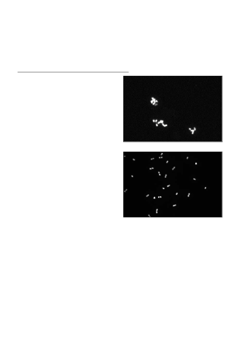
visualized under blue-green light excitation (488 nm). Results are
shown in figures 1 and 2; data obtained showed the successful ampli-
fication of 16S rDNA in whole A. venetianuscells (Fig. 1), whereas no
?uorescent signal was found in the control experiment (Fig. 2). The
same results were obtained with E. coliXl1-blue cells (not shown).
In order to check the possibility of using other target sequences for
in-situ- amplification, we choose the A. venetianus gyrBsequence
(encoding the b-subunit of DNA gyrase) as target in the further exper-
iments. For this purpose, the VE-C3 gyBsequence was firstly ampli-
fiedviaPCR by using the primers previously described by Yamamoto
et al (4). The amplification product was cloned, and its nucleotide
sequence determined.
Two oligonucleotides were then designed on this sequence and used
as primers in in-situPCR experiments on whole A. venetianusVE-C3
cells. Data obtained (not reported) revealed the amplification of the
gyrBsequence inside the A. venetianuscells.
This body of data suggested that in-situ PCR can be successfully
used to amplify DNA sequences within Acinetobatercells and might
represent a powerful tool for identification and monitoring of this bac-
terium in microbial communities.
References
1. Jacobs D, Ales ML, Goodman AE and Neilan BA (1997). Improved
methods for in-situenzymatic amplification and detection of low copy
number genes in bacteria. FEMS Microbiol. Letters, 152: 65-73.
2. Hodson RE, Dustman WA, Garg RP and Moran MA (1995). In-situ
PCR for visualization of microscale distribution of specific genes and
gene products in prokaryotic communities. Appl. Environ. Microbiol., 61:
4075-4082.
3. Chen F, Gonzales JM, Dustman WA, Moran MA and Hodson RE
(1997).In-situreverse trascription, an approach to charaterize genetic
diversity and activities of Prokaryotes. Appl. Environ. Microbiol., 63:
4907-4913.
4.Yamamoto S, Bouvet PJM and Harayama S (1999). Phylogenetic
structures of genus Acinetobacterbased on gyrBsequences: comparison
with the grouping by DNA-DNA hybridization. International Journal of
Systematic Bacteriol., 49 87-95.
5. Di Cello F, Pepi M, Baldi F and Fani R (1997). Molecular
characterization of an n-alkane degrading bacterial community and
identification of a new species, Acinetobacter venetianus. Res.
Microbiol., 148: 237-249.
Rapp. Comm. int. Mer Médit., 36,2001
IN SITUPCR AMPLIFICATION OF 16SRDNAANDGYRBGENE IN ACINETOBACTER VENETIANUSCELLS
M. Betti
1
, F.Torricelli
2
, S. Bonifacio
2
, F. Baldi
3
, M. Pepi
3
and R. Fani
1
*
1
Dipartimento di biologia animale e genetica, Universitŕ degli Studi, Firenze, Italy
2
Citogenetica e genetica U.O., A.O. Careggi, Firenze, Italy
3
Dipartimento di Scienze ambientali, Venezia, Italy - r_fani@dbag.unifi.it
Abstract
Thein-situpolymerase chain reaction (in-situPCR) is a molecular technique combining the extreme sensitivity of the PCR with the in-
situhybridization.In-situPCR is used to detect very low quantities of DNA in intact cells. We present a protocol, which has been used to
amplify the 16S rDNA and the gyrBgene in Escherichia coliand inAcinetobacter venetianuscells. The latter has been isolated from the
Venice Lagoon and is able to perform the efficient oxidation of n-hydrocarbons and could be used for bioremediation purposes in polluted
areas.
Keywords: in-situ PCR, Acinetobacter venetianus, Escherichia coli, FISH.
Fig. 1. In-situPCR amplification of 16S rDNAsequence in VE-
C3Acinetobacter venetianusfixed cells.
Fig. 2.In-situPCR control reactions.